| dc.contributor.author | Onder, Sevda | |
| dc.contributor.author | Bilgili, Serap Gunes | |
| dc.contributor.author | Bulut, Gulay | |
| dc.contributor.author | Celik, Huseyin Tugrul | |
| dc.contributor.author | Oguztuzun, Serpil | |
| dc.contributor.author | Onder, Haci | |
| dc.contributor.author | Calka, Omer | |
| dc.date.accessioned | 2021-01-14T18:11:01Z | |
| dc.date.available | 2021-01-14T18:11:01Z | |
| dc.date.issued | 2020 | |
| dc.identifier.citation | Önder, S., Güneş Bilgili, S., Bulut, G., Celik, H. T., Oguztuzun, S., Onder, H., … Karadağ, A. S. (2020). Biochemical and histopathologic assessment of effects of acitretin on epiphyseal growth plate in rats. Advances in Dermatology and Allergology, 37(3), 346–352. | en_US |
| dc.identifier.issn | 1642-395X | |
| dc.identifier.uri | https://doi.org/10.5114/ada.2020.95983 | |
| dc.identifier.uri | https://hdl.handle.net/20.500.12587/12854 | |
| dc.description | WOS:000551559000009 | en_US |
| dc.description | PubMed: 32792874 | en_US |
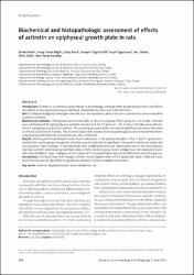
| dc.description.abstract | Introduction: Acitretin is a commonly used retinoid in dermatology. Although there are generally known side effects, the effects on the epiphyseal plaque and bone metabolism are not clear in the literature. Aim: To histopathologically investigate the effects on the epiphyseal plate and assess variations in bone metabolism caused by acitretin. Material and methods: Three groups were formed with 10 rats in each group. The 1st group (n = 10, 5 male, 5 female) were administered 10 mg/kg/day oral acitretin solution and the 2nd group (n = 10, 5 male, 5 female) were administered 3 mg/kg/day oral acitretin solution. The control group were given normal standard feed and water. Rats were sacrificed at the end of 4 weeks. The proximal tibias were excised and histopathologically and immunohistochemically assessed. Biochemical assessment was also carried out. Results: Staining with haematoxylin-eosin found reductions in the epiphyseal plate in the 1st and 2nd group compared to the control group, though this situation was not statistically significant. Immunohistochemical studies did not encounter Type II collagen in the epiphyseal bone, proliferative zone and hypertrophic zone in the control group, low dose acitretin solution group and high dose acitretin solution group. Type II collagen was not observed in osteoids and osteoblasts. Type I collagen was not observed in the hypertrophic zone and proliferative zone of any group. Conclusions: Our data show that though acitretin caused degeneration of the epiphyseal plate, it did not cause clear thinning and we identified no significant variations in bone metabolism markers. | en_US |
| dc.description.sponsorship | Yuzuncu Yil University Scientific Research Project Chair | en_US |
| dc.description.sponsorship | We thank the Yuzuncu Yil University Scientific Research Project Chair for funding this research.; This study was conducted in the Department of Dermatology of the Yuzuncu Yil University. | en_US |
| dc.language.iso | eng | en_US |
| dc.publisher | TERMEDIA PUBLISHING HOUSE LTD | en_US |
| dc.relation.isversionof | 10.5114/ada.2020.95983 | en_US |
| dc.rights | info:eu-repo/semantics/openAccess | en_US |
| dc.subject | acitretin | en_US |
| dc.subject | epiphyseal plate | en_US |
| dc.subject | bone metabolism | en_US |
| dc.subject | rat | en_US |
| dc.title | Biochemical and histopathologic assessment of effects of acitretin on epiphyseal growth plate in rats | en_US |
| dc.type | article | en_US |
| dc.contributor.department | KKÜ | en_US |
| dc.identifier.volume | 37 | en_US |
| dc.identifier.issue | 3 | en_US |
| dc.identifier.startpage | 346 | en_US |
| dc.identifier.endpage | 352 | en_US |
| dc.relation.journal | POSTEPY DERMATOLOGII I ALERGOLOGII | en_US |
| dc.relation.publicationcategory | Makale - Uluslararası Hakemli Dergi - Kurum Öğretim Elemanı | en_US |