| dc.contributor.author | Gültiken, Murat Erdem | |
| dc.contributor.author | Yıldız, Dinçer | |
| dc.contributor.author | Karahan, Siyami | |
| dc.contributor.author | Bolat, Durmuş | |
| dc.date.accessioned | 2021-01-14T18:19:40Z | |
| dc.date.available | 2021-01-14T18:19:40Z | |
| dc.date.issued | 2010 | |
| dc.identifier.citation | GÜLTİKEN M. E,YILDIZ D,KARAHAN S,BOLAT D (2010). Scanning electron and light microscopic investigation of Bursa fabricius in turkey (Meleagris gallopavo). Eurasian Journal of Veterinary Sciences, 26(2), 69 - 73. | en_US |
| dc.identifier.issn | 1309-6958 | |
| dc.identifier.issn | 2146-1953 | |
| dc.identifier.uri | https://app.trdizin.gov.tr/makale/TVRBNU5qVXpNdz09 | |
| dc.identifier.uri | https://hdl.handle.net/20.500.12587/13790 | |
| dc.description.abstract | Gültiken ME, Yıldız D, Karahan S, Bolat D. Hindi (Meleagris gallopavo) Bursa fabricius’unun taramalı elektron ve ışık mikroskobu ile incelenmesi. Eurasian J Vet Sci, 2010, 26, 2, 69-73 Amaç: Hindide Bursa fabricius’un morfolojisinin belirli dönemlerde incelenmesi ve dönemlere göre morfometrik analizinin taramalı elektron mikroskobu ve ışık mikroskobu kullanılarak yapılmasıdır. Gereç ve Yöntem: Çalışmada 1-24 haftalığa kadar toplam 50 hindiye ait Bursa fabricius’lar kullanıldı. Hayvanların ve nekropsi sonrası Bursa fabricius’ların ağırlıkları belirlendi. Taramalı elektron mikroskobu ile yapılacak çalışma öncesi dokular gluteraldehit ile tespit edildikten sonra mikroskop kullanılarak görüntüler elde edildi. Histolojik çalışma için dokular rutin histolojik takip sonrası Mallory’s triple ve Haematoxylen-eosin ile boyanarak ışık mikroskobu ile incelendi. Bulgular: Morfometrik veriler Bursa fabricius’un maksimum büyüklüğüne 9. haftada ulaştığını gösterdi. Dokuzuncu haftayı takiben Bursa fabricius ağırlığındaki azalma hindide involusyonun bu dönemi takiben şekillenmeye başladığını gösterdi. Taramalı elektron mikroskop ile yapılan incelemelerde Bursa fabricius lumenine uzanan plica yüzeylerindeki kubbe şeklindeki epitel görüntüsünün 5. ve 9. haftalarda en belirgin ve düzgün olarak tespit edildi. İlerleyen haftalarda (13. ve 24.) Bu manzarada dikkat çekici bir düzensizlik gözlendi. Öneri: Çalışma sonucunda elde edilen bulguların, ilerde yapılacak çalışmalara ve hindi aşılama programlarına katkı yapacağı düşünülmektedir. | en_US |
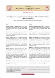
| dc.description.abstract | Gultiken ME, Yildiz D, Karahan S, Bolat D. Scanning electron and light microscopic investigation of Bursa fabricius in turkey (Meleagris gallopavo). Eurasian J Vet Sci, 2010, 26, 2, 69-73 Aim: The aim of this study was postnatal investigation of morphometric features of Bursa fabricius in Turkey by using scanning electron microscope and light microscope. Materials and Methods: One 1-24 week old 50 turkeys (meleagris gallopavo) were used. Their Bursa fabriciuses were taken out after the necropsy and weighted. Tissues were fixed with glutaraldehyde, examined and photographed under scanning electron microscope. For histological examination, tissues were prepared using routine histologic methods and stained with Mallory’s triple and Hematoxylen-Eosine. Results: The morphometric data concerning turkey ducklings used in the study showed that Bursa fabricius reaches its maximum size at the 9th week. The decrease in the weight of Bursa fabricius following the week of 9 proved that involution session in the turkey begins after that period. On the investigation performed by means of scanning electron microscopy, dome shaped surface epithelium which covers subepithelial lymph follicles on the surface was determined to be most clear in the 5th and 9th weeks. There was a distinct irregularity of this appearance in the following weeks ($13^ {th}$ and $24^ {th}$). Conclusion: It was concluded that the results may contribute to the researches which relate to humoral immunity formation and effectiveness of vaccination programme. | en_US |
| dc.language.iso | eng | en_US |
| dc.rights | info:eu-repo/semantics/openAccess | en_US |
| dc.subject | Veterinerlik | en_US |
| dc.title | Scanning electron and light microscopic investigation of Bursa fabricius in turkey (Meleagris gallopavo) | en_US |
| dc.title.alternative | Hindi (Meleagris gallopavo) Bursa fabricius’unun taramalı elektron ve ışık mikroskobu ile incelenmesi. | en_US |
| dc.type | article | en_US |
| dc.identifier.volume | 26 | en_US |
| dc.identifier.issue | 2 | en_US |
| dc.identifier.startpage | 69 | en_US |
| dc.identifier.endpage | 73 | en_US |
| dc.relation.journal | Eurasian Journal of Veterinary Sciences | en_US |
| dc.relation.publicationcategory | Makale - Ulusal Hakemli Dergi - Kurum Öğretim Elemanı | en_US |


