| dc.contributor.author | Say, Bahar | |
| dc.contributor.author | Yıldız, Ayşe | |
| dc.contributor.author | Alpua, Murat | |
| dc.contributor.author | Ergun, Ufuk | |
| dc.date.accessioned | 2021-01-14T18:22:00Z | |
| dc.date.available | 2021-01-14T18:22:00Z | |
| dc.date.issued | 2020 | |
| dc.identifier.citation | Say, B., Yıldız, A., Alpua, M., Ergun, U. (2020). Electroencephalography (EEG) and Syncope: A Retrospective Study. Epilepsi, 26(2), 103 - 107. | en_US |
| dc.identifier.issn | 1300-7157 | |
| dc.identifier.uri | https://doi.org/10.14744/epilepsi.2020.36036 | |
| dc.identifier.uri | https://app.trdizin.gov.tr/makale/TXpnek9UUTRPQT09 | |
| dc.identifier.uri | https://hdl.handle.net/20.500.12587/13975 | |
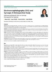
| dc.description.abstract | Objectives: Electroencephalography (EEG) is one of the most vital tools in neurology practice. It is used for the diagnosis and differential diagnosis of several clinical conditions. One of them is syncope. In this study, it was planned that a retrospective evaluation of EEGs performed due to syncope in our laboratory and determine the rate of abnormal EEGs. Methods: EEG recordings performed due to syncope were reviewed over a two-years period in this study. The EEG findings were classified as normal and abnormal. The abnormal EEG findings were classified into focal epileptiform discharge, generalized epileptiform discharge, focal slowing and generalized slowing subgroups and analyzed. Results: The results of 298 EEGs were analyzed, which involved 174 (58.3%) female and 124 (41.6%) male subjects, with a mean age of 38.84±17.83 (min-max: 17–90) years. Among subjects with syncope, 90.6% of the EEGs were normal and 9.4% showed abnormal findings. The most common abnormal finding was focal epileptiform discharge (5.03%). Generalized epileptiform discharge was observed in three (1%), focal slowing in seven (2.3%) and generalized slowing in two (0.6%) subjects. Among the EEG results with no abnormal findings, 38 (12.7%) had sleep-deprived EEG and six (2.1%) were found to have focal epileptiform discharge. A total of 113 (37.9%) subjects had electrocardiogram recording and results were normal. Conclusion: The rate of abnormal findings in EEGs performed due to syncope is low. EEG may be helpful in some selected subjects with syncope referred to the laboratory with a good history and neurological examination. EEG may be repeated if necessary | en_US |
| dc.description.abstract | Amaç: Elektroensefalografi (EEG) nöroloji pratiğinde önemli yere sahip araçlardan biridir. Birçok klinik durumun tanı ve ayırıcı tanısında kullanılabilmektedir. Bunlardan biri de senkoptur. Bu çalışmada, senkop nedeniyle laboratuarımızda çekilen EEG’lerin retrospektif değerlendirilmesi ve anormal EEG oranının belirlenmesi planlandı. Gereç ve Yöntem: Senkop nedeniyle çekilen iki yıllık EEG kayıtları retrospektif olarak gözden geçirildi. Olguların EEG sonuçları normal (anormal bulgu içermeyen) ve anormal olarak sınıflandırıldı. Anormal olan EEG sonuçlarında fokal epileptiform deşarj, jeneralize epileptiform deşarj, fokal yavaş, jeneralize yavaş olarak alt gruplara bakıldı. Bulgular: Bu çalışmaya 298 EEG dahil edildi. Çalışmada 174 (%58.3) kadın, 124 (%41.6) erkek olup yaş ortalaması 38.84±17.83 (min-maks: 17–90) oldu. Senkoplu olgular arasında EEG’lerin %90,6’sı normal, %9.4’ü anormaldi. En sık fokal epileptiform deşarj (%5.03) izlendi. Jeneralize epileptiform deşarj üç (%1), fokal yavaşlama yedi (%2.3), jeneralize yavaşlama iki (%0.6) olguda izlendi. Anormal bulgu içermeyen EEG’ye sahip olguların 38’ine (%12,7) uyku deprivasyonlu EEG çekilmişti ve altısında (%2.1) fokal epileptiform deşarj saptanmıştı. Toplamda 113 (%37.9) olguda eş zamanlı elektrokardiografi kayıdı vardı ve sonuçları normaldi. Sonuç: Senkop nedeniyle çekilen EEG’lerde anormal bulgu oranı düşüktür. İyi bir öykü ve nörolojik muayene ile laboratuara yönlendirilen seçilmiş bazı senkop olgularında EEG faydalı olabilir. Gerekirse EEG tekrar edilebilir. | en_US |
| dc.language.iso | eng | en_US |
| dc.relation.isversionof | 10.14744/epilepsi.2020.36036 | en_US |
| dc.rights | info:eu-repo/semantics/openAccess | en_US |
| dc.title | Electroencephalography (EEG) and Syncope: A Retrospective Study | en_US |
| dc.title.alternative | Elektroensefalografi (EEG) ve Senkop: Retrospektif Bir Çalışma | en_US |
| dc.type | article | en_US |
| dc.identifier.volume | 26 | en_US |
| dc.identifier.issue | 2 | en_US |
| dc.identifier.startpage | 103 | en_US |
| dc.identifier.endpage | 107 | en_US |
| dc.relation.journal | Epilepsi | en_US |
| dc.relation.publicationcategory | Makale - Ulusal Hakemli Dergi - Kurum Öğretim Elemanı | en_US |


